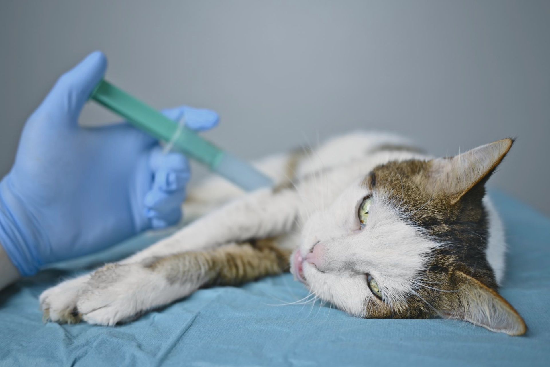
Feline panleukopaenia
Feline
panleukopenia is a highly contagious viral disease that affects cats,
particularly in communities. Prevalence data is limited, but there may have
been an increase in cases in certain areas. It is caused by feline parvovirus
(FPV), a contagious DNA virus that affects all members of the Felidae family.
Control of the disease is complicated due to environmental resistance, high
viral shedding, and interspecies transmission.
The
severity of clinical signs varies based on the cat's immune status and
concurrent diseases. Symptoms range from mild or no apparent signs to a
hyperacute syndrome that can result in sudden death. Common clinical signs
include fever, lethargy, anorexia, vomiting, and watery or bloody diarrhea.
Secondary bacterial infections can lead to complications such as sepsis,
dehydration, and disseminated intravascular coagulation.
Some
parvoviruses, like canine parvovirus type 1 (CPV-1) and FPV, have undergone minimal
evolution over time. However, canine parvovirus type 2 (CPV-2) has shown
significant adaptability and mutation, leading to the development of three
pathogenic strains: CPV-2a, CPV-2b, and CPV-2c.
The
United States has seen a significant increase in FPV cases, possibly due to
changes in vaccination protocols. The use of intranasal vaccines has risen to
mitigate the risk of vaccine-associated sarcomas. However, these vaccines may
be less effective, especially in high-risk animals, compared to injectable
vaccines. New vaccination guidelines recommend completing primary vaccination
at 14 to 16 weeks of age based on studies showing extended persistence of
maternal antibodies. It is possible that these new protocols are not yet widely
adopted by most veterinary surgeons.
There is a lack of published data on the prevalence increase of feline panleukopenia in Spain. However, based on experience, there has been an increase in diagnosed cases in recent years, with 195 cases reported in a span of six years.
Aetiology
Feline panleukopenia virus (FPV) is a
small non-enveloped DNA virus, measuring 20 nm in size. It is primarily
transmitted through the faecal-oral route, and infected cats intermittently shed
the virus in their feces for days or weeks.
The virus is highly contagious and
can be easily transmitted through various means, including clothing, shoes,
utensils, and even flies.
Disinfecting contaminated areas is
challenging due to the virus's resistance to chemical disinfectants and
physical agents. Effective disinfectants against FPV include sodium
hypochlorite, formaldehyde, and glutaraldehyde.
The replication of the virus begins
in the oropharyngeal tissue. As a single-stranded DNA virus, FPV requires
actively dividing cells in the S phase. Additionally, the virus relies on DNA
polymerase enzyme to synthesize the complementary DNA strand, making it
dependent on tissues with high mitotic activity. After replication, the virus
enters the bloodstream, leading to viremia and dissemination to target organs
such as tonsils, mesenteric lymph nodes, lymphatic intestinal tissue, and bone
marrow. The pathogenic consequences and clinical manifestations of FPV vary
depending on the specific tissue infected (see Table 1 below).
Table 1. Pathogenic consequences and clinical manifestations of FPV
depending on the tissue infected.
|
Infected tissue |
Consequences |
Clinical manifestations |
|
Intestinal
crypt epithelium |
Collapse of villi |
Enteritis |
|
Lymph nodes and thymus |
Lymphocyte apoptosis, thymic atrophy |
Lymphopaenia |
|
Bone
marrow |
Decrease in stem cell number |
Pancytopaenia |
|
Foetus |
Foetal death |
Abortion |
|
Developing
cerebellum |
Cerebellar hypoplasia |
Ataxia |
Pathogenesis and Clinical Signs
The incubation period of feline
panleukopenia, from virus exposure to the onset of clinical signs, ranges from
5 to 9 days. Four types of clinical manifestations are observed.
1. Subclinical infection: This is the most common outcome, where infected cats show no
apparent clinical signs of the disease. The development of clinical disease
depends on various factors, including the rate of intestinal crypt cell
mitosis. Low rates of mitosis result in minimal intestinal damage, while high
rates, triggered by stress or concurrent diseases, provide an optimal
environment for the virus to replicate and cause more severe effects. Even
dietary changes can increase the rate of mitosis, facilitating viral
replication. For example, adopted kittens may develop severe enteritis and die,
while littermates remaining with the mother or in a shelter may show milder or
no clinical signs.
2. Sudden death: Sudden death is relatively common, especially in young
kittens, unvaccinated cats, and animals in shelters or pet shops.
3. Classic feline enteritis: This clinical presentation typically occurs suddenly.
Owners may mistake the signs for poisoning. Clinical signs include fever,
anorexia, dehydration, vomiting, and, in some cases, bloody diarrhea.
Pancytopenia (reduced blood cell counts) is often observed in complete blood
counts. Severe dehydration and loss of the intestinal barrier, leading to
bacterial overgrowth, can result in death.
4. Neurological signs: The virus's preference for dividing cells makes the fetus
highly vulnerable. Infection during the first trimester of gestation can cause
abortion, fetal resorption, and death. Infections at later stages can lead to cerebellar
hypoplasia, hydrocephalus with ataxia, intention tremors, and limb
abnormalities.
Laboratory Diagnosis
Laboratory diagnosis of feline
panleukopenia involves several methods:
1. Virus isolation: The virus can be isolated from stool samples, although this
can be challenging.
2. Antigen detection: Rapid immunochromatographic tests used for detecting canine
parvovirus (CPV) antigen can also be used for FPV detection. However, the
results should be interpreted carefully, as the test may cross-react with
vaccinated animals and yield false positives.
3. PCR: Polymerase chain reaction (PCR) can detect viral genetic material (DNA)
in blood or stool samples. Blood samples are preferred when the animal has
diarrhea.
4. Antibody detection: ELISA and indirect immunofluorescence (IF) are commonly used
methods to detect serum antibodies. Differentiating between vaccine-induced
antibodies and infection-induced antibodies can be challenging, particularly in
vaccinated animals.
Treatment
If a cat is hospitalized with a
confirmed or suspected diagnosis of feline panleukopenia, strict isolation
measures should be implemented to prevent virus spread.
Supportive care for clinical signs includes:
1. Fluid therapy: Intravenous fluid therapy and electrolyte control are
crucial. Central venous catheterization is preferred for prolonged
hospitalization to manage hypoalbuminemia and peripheral edema.
2. Antibiotic therapy: Broad-spectrum antibiotics are administered to prevent
bacterial infections due to the compromised intestinal barrier. A combination
of amoxicillin and clavulanate with a third-generation cephalosporin is an
option.
3. Parenteral nutrition: In some cases, supplemental nutrition may be necessary.
4. Antiemetics: Medications to control vomiting and alleviate nausea and
discomfort.
5. Transfusions: Whole blood or plasma transfusions may be required.
6. Analgesics: Acute pain management is essential, and opioids such as
buprenorphine or fentanyl can be used.
Specific Antiviral Therapy
1. Antiviral chemotherapy: Feline
interferon ω can be administered intravenously at a dose of 2.5 MIU/kg every 24
hours for three consecutive days. Limited studies exist, with a higher level of
scientific evidence needed.
2. Passive immunotherapy:
Subcutaneous administration of serum or homologous plasma from recovered or
vaccinated animals with high antibody titers can be considered. Blood group
compatibility should be determined beforehand. High-quality studies supporting
this approach are lacking.
Prevention
Vaccination:
Vaccination is the most effective
method of preventing feline panleukopenia. Live attenuated vaccines are used,
starting at 8 to 9 weeks of age, with doses administered every 3 weeks
until the final vaccine is given after 16 weeks of age. Revaccination
should be done after 1 year. It is important to vaccinate all cats, even those
that do not go outdoors.
Maternal antibodies are transferred
to kittens through colostrum. Kittens should nurse within the first 8 hours of
life to acquire these antibodies. Maternal antibody levels decline from the
10th week of life. Vaccinating too early can interfere with active immunity due
to passive immunity from maternal antibodies. Recommendations for different
scenarios include vaccinating at 8 to 9 weeks of age for kittens that nursed
well, vaccinating early (7-12 days of age) with an inactivated vaccine for
kittens that did not nurse within the first hours of life, and vaccinating
gestating cats with inactivated vaccines. Retrovirus-positive animals should
receive inactivated vaccines if their health permits.
Live attenuated vaccines provide
immunity within three days of vaccination. A general vaccination schedule
includes vaccines at 8-9 weeks, 12 weeks, and after 16 weeks of age, with
revaccination after 1 year.
Sanitary Period:
Implementing an adequate sanitary
period is crucial for eradicating the virus, especially in feline communities.
The virus can persist in the environment for up to a year. Disinfection using
sodium hypochlorite (diluted bleach) can reduce viral load. Establishing
contamination-free areas in high-risk places such as shelters, catteries,
foster homes, and veterinary clinics is recommended.
Hyperimmune Sera:
Hyperimmune sera can be administered
before a high-risk situation, but there is a lack of scientific evidence
demonstrating its effectiveness.
Feline Interferon ω:
A study conducted in naturally
infected cats found no significant differences in survival rate or clinical
signs between cats pretreated with interferon and untreated cats.
Prognosis:
In cats infected with feline
panleukopenia, leukopenia, lymphopenia, monocytopenia, and eosinopenia indicate
a poor prognosis. Decreased cortisol, thyroxine, and serum cholesterol
concentrations further complicate the prognosis. Systemic inflammatory response
syndrome (SIRS) is more common in dogs that do not survive and have concurrent
viral, bacterial, or parasitic diseases. Although data for feline panleukopenia
is limited, further studies are necessary to improve treatment, anticipate
complications, and enhance survival rates.
Results of a Retrospective Study:
Findings of a retrospective include:
- The survival rate was 52%, with
most infections occurring during the first year of life, particularly between 3
and 4 months of age. However, FPV was also diagnosed in adult cats and those
without outdoor access.
- Cats infected with FPV had received
one or two vaccinations, but none had completed the full vaccination schedule
or received vaccines after 16 weeks of age.
- Common clinical signs at diagnosis
included anorexia, lack of energy, vomiting, and diarrhea.
- Cats with concomitant respiratory
infections had a higher mortality rate.
- Significant laboratory findings
included leukopenia and hypoalbuminemia.
Frequent Questions
1. Can a dog infect a cat?
Cats can be infected by both feline
parvovirus (FPV) and canine parvovirus (CPV-2a, CPV-2b, and CPV-2c). Both
viruses can cause panleukopenia-like signs. Test kits designed for detecting
canine parvovirus antigens can also detect both wild and vaccine strains of
FPV.
2. Can a cat infect a dog?
The ability of a cat to infect a dog
depends on the strain involved. Cats shedding CPV-2a or CPV-2b antigen can
infect dogs, but cats shedding FPV cannot infect dogs.
3. If the result of the antigen test is negative, can I rule out FPV
infection?
Even if the result of the fecal
antigen test is negative, FPV infection cannot be ruled out. Viral shedding can
occur intermittently or over a short period, and the test may yield false
negatives if performed 5 to 7 days after the onset of disease. Viremia occurs
before fecal shedding, so a negative result can be obtained if the test is
performed too soon. In the early stages of the disease, PCR analysis of blood
or bone marrow can provide an earlier positive result for FPV compared to the
fecal antigen test.
4. Can an indoor cat become infected?
Yes, an indoor cat can be indirectly
exposed to the virus through contaminated shoes, bags, or objects that serve as
vehicles for disease transmission, carrying either canine or feline parvovirus.
5. Can a vaccinated cat become infected with FPV?
Yes, if the complete vaccination
schedule has not been followed. The most common cause of disease in vaccinated
cats is interference from maternal antibodies. Vaccination should begin at 9
weeks and 12 weeks, with the final dose always administered after 16 weeks.
These new vaccination recommendations are supported by experts in feline
infectious diseases.
6. Why are neurological signs observed in kittens infected with FPV?
Kittens infected with FPV can develop
cerebellar disease, which is characterized by symmetrical ataxia, hypermetric
movements of the limbs and head, and intention tremors. This is a result of
cerebellar hypoplasia caused by intrauterine FPV infection. Affected kittens
usually come from litters where multiple kittens are affected, leading to
confusion with congenital heart disease. However, affected kittens present with
non-progressive cerebellar ataxia once they start walking.
7. How is cerebellar hypoplasia diagnosed? Is it contagious to other cats?
Diagnosis of cerebellar hypoplasia
can be made through magnetic resonance imaging (MRI) or post-mortem analysis,
but there is no specific test for detecting parvovirus due to the absence of an
active infection. Cerebellar hypoplasia is not contagious to other cats.
Affected cats gradually adapt to neurological deficits and can lead normal
lives.
8. What vaccination schedule is recommended for an adult cat with unknown
vaccination status?
It is highly recommended to vaccinate
the cat with two vaccinations separated by 3 weeks.
9. What is the most appropriate treatment? Is antibiotic use appropriate?
Cats with feline panleukopenia should
be isolated and provided with intensive treatment. Intravenous fluid therapy is
crucial, correcting metabolic acidosis and hypokalemia. Restricting food and
water intake should only be done if there is vomiting, and it should be
restored as soon as possible. Symptomatic medications can be administered for
vomiting or nausea. Transfusion of whole blood or plasma may be necessary in
cases involving hypoproteinemia. Antibiotics specific to gram-negative and
anaerobic bacteria should be given intravenously to prevent bacterial
contamination due to intestinal barrier compromise. Treatment should be
tailored to individual cases.
10. What should be done in case of an outbreak of FPV at a clinic?
In case of an outbreak, it is
important to implement cleaning and disinfection measures as part of the
infectious disease control protocol. Disinfectants should be used after washing
biological material with soap. Unvaccinated animals should be handled with
gloves and placed on clean surfaces. Surfaces and objects that are not
routinely cleaned, such as doorknobs, keyboards, light switches, and
countertops, should be periodically cleaned. Personnel handling sick animals
should use gloves, gowns, and foot coverings, and a color code can be used to
mark contaminated material. Knowledge of the vaccination status of other
animals attending the clinic is crucial.
Key Points:
- Kittens are highly susceptible to
FPV infection before acquiring vaccine antibodies, following a decline in
maternal antibody levels.
- Vaccination against FPV is
recommended, tailored to each animal's circumstances, with vaccination after 16
weeks of age.
- Adult and indoor cats are also
susceptible to FPV infection.
- Clinical presentation can vary,
with anorexia and depression being common signs without vomiting or diarrhea.
- Rapid tests provide a reliable
diagnosis, but false negatives are possible. Results should be interpreted
considering the clinical presentation and the animal's risk of exposure. PCR
analysis can be used to verify diagnosis.
- Hospitalization and supportive
treatment are essential. The duration of hospitalization ranges from 5 to 9
days.
- Concomitant diseases worsen the
prognosis, regardless of the animal's age.
- Enteral nutrition is beneficial for
vomiting cases, while parenteral or microenteral nutrition can be alternatives.
Hypoalbuminemia and leukopenia may be negative prognostic factors.
- Nursing care, keeping cats warm,
dry, and clean, is crucial.
- There is a high risk of hospital
contamination.
Case Study 1: Ketty
Summary: Ketty is a 5-month-old
non-neutered male cat. He was adopted from a shelter 7 days prior to the
consultation. The reason for the consultation is weakness and anorexia.
Medical History:
Ketty was vaccinated with an
inactivated vaccine against feline panleukopenia virus (FPV), feline
herpesvirus (FHV), and feline calicivirus (FCV) at 8 weeks of age. Since
arriving at his new home, Ketty has been hiding and it is unclear if he has
been eating properly. The owners assumed his behavior was normal for a cat in a
new environment. However, Ketty was active and friendly during his time at the
shelter.
Physical Examination:
Ketty's physical examination reveals
a body condition of 2/5, weighing 1.7 kg. He shows weakness, lethargy,
dehydration (approximately 7%), excessive salivation, abdominal pain on
palpation, rectal temperature of 40.9 °C, bradycardia (60 beats per minute),
and a capillary refill time >2 seconds. A complete blood count is performed.
Diagnoses:
The most likely diagnosis for Ketty
is feline panleukopenia virus (FPV) due to his clinical signs, exposure to a
risky environment, inadequate vaccination history (not vaccinated after 16
weeks), and severe leukopenia observed in the complete blood count.
Treatment:
Ketty requires complete isolation and
hospitalization with intensive care. Fluid replacement therapy and
antimicrobial treatment are administered. Placement of a central venous
catheter is advised if possible. Enteral nutrition is initiated after
supportive treatment. Ketty shows significant improvement and is discharged.
Case Study 2: Kitty and his two littermates
Review: Kitty and his two littermates
are domestic shorthair cats, 2 months old, neither vaccinated nor dewormed. The
reason for consultation is a lack of energy.
Medical History:
Kitty and his littermates were found
on the street and appear to be from the same litter. They have been in their
new home for 10 days and initially showed normal eating and playing behavior.
However, Kitty stopped playing and eating three or four days before the
consultation.
Physical Examination:
Kitty's physical examination reveals
a body condition of 3/5, weighing 200 g (lower than his littermates). He
displays weakness, lethargy, poor coat, dehydration (approximately 8%),
excessive salivation, abdominal pain on palpation, rectal temperature of 41.2
°C, bradycardia (80 beats per minute), and a capillary refill time >2
seconds. A complete blood count is performed, and a fecal ELISA for canine
parvovirus antigen produces a positive result.
Treatment:
Kitty needs to be hospitalized in
isolation due to the hyperacute clinical signs and positive diagnosis of FPV.
His littermates should be monitored and potentially undergo additional tests.
In addition to fluid therapy and antibiotic treatment, a blood transfusion and
parenteral nutrition are proposed.
Evolution:
After receiving a blood transfusion and parenteral nutrition, Kitty starts receiving enteral nutrition. He experiences complications with an upper airway respiratory process, requiring extended hospitalization. After 7 days of hospitalization with fluid and supportive therapy, Kitty's body weight increases to 270 g, and he makes a full recovery. One of his littermates dies suddenly, and necropsy reveals intestinal lesions consistent with FPV. The third littermate shows no signs of disease or hematological disorders.


